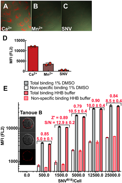
Figure 1.
Confocal microscopy images show differential binding of SNVR18 to Vero E6 cells in HHB/0.1% HSA buffer in the presence of A. 1.5 mM Ca2+ which supports an integrin bent conformation low-affinity state, B. 1 mM Mn2+ activates integrins and induces an extended integrin conformation. C. Excess unlabeled SNV used to block SNVR18. D. Flow cytometry measurements of SNVR18 binding to Vero E6 cells in the presence Ca2+, Mn2+, and excess unlabeled SNV. 100,000 Vero E6 /well, were incubated with 1500 SNVR18/cell at 37°C under mild vortexing for 15 mins. The cells were then washed 3x in the buffer, then either removed from the bottom of the wells by 0.025% Trypsin/0.0526 mM EDTA for flow cytometric measurement or fixed in 2% PFA at 0°C for 30 mins before mounting in Vector Shield containing 1X DAPI for microscopic imaging. E. Equilibrium binding of SNVR18 titers to 10,000 Tanoue B cells in a 384 well plate. Quadruplicate measurements were performed for each condition to determine the signal to background ratio and Z’ values for each titer. The Z’-score values for each condition were comparable, here Z’ scores for +DMSO are shown. Error bars represent ±SD.

