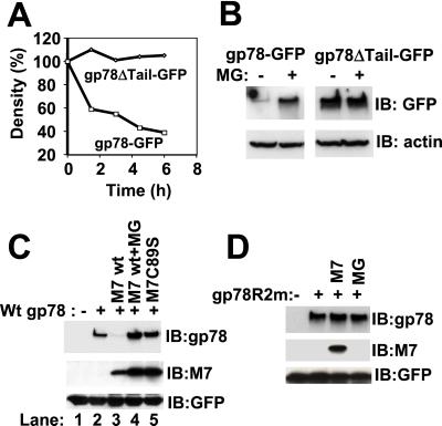
Figure 5.
RING-, proteasome-, and MmUBC7-dependent degradation of gp78. (A) Cells stably expressing GFP-tagged wt gp78 (gp78-GFP) or GFP-tagged gp78 lacking the C-terminal tail (gp78ΔTail-GFP) were pulse labeled with [35S]methionine for 30 min followed by removal of unincorporated [35S]methionine. gp78 was then immunoprecipitated by using anti-GFP, and, after resolution on SDS/PAGE, gp78 was quantified and plotted as a function of the amount at the initiation of the chase. (B) Cells stably expressing gp78-GFP or gp78ΔTail-GFP were treated with or without MG132 (MG) for 6 h followed by immunoblotting with anti-GFP; β-actin was used as a gel loading control. (C and D) Lysates from cells transfected as indicated were processed for immunoblotting for gp78, MmUBC7 (M7), and cotransfected GFP (transfection efficiency control). Cells exposed to MG132 (MG) were treated with 20 μM for 6 h.

