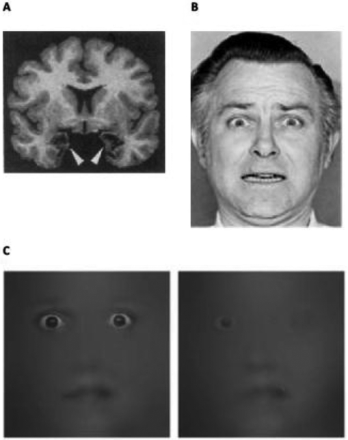
Figure 1. Perceiving Fear Without An Amygala.
(A) Tl-weighted magnetic resonance (MR) image of S.M.’s brain in the coronal plane, showing amygdala lesions on the left and right (dark regions, denoted by arrows), from [4] with permission. S.M.’s lesions are largely confined to her amygdalae, and the fibers of passage therein (although additional damage was later detected in the anterior portion of her entorhinal cortex, the adjacent white matter, and a portion of her ventromedial prefrontal cortex; [1]). (B) An example of a posed, wide-eyed gasping face that symbolizes fear. (C) Facial information used to discriminate a wide-eyed gasping face from a smiling face in ten control subjects (left) and S.M (right) (from [10], used with permission).

