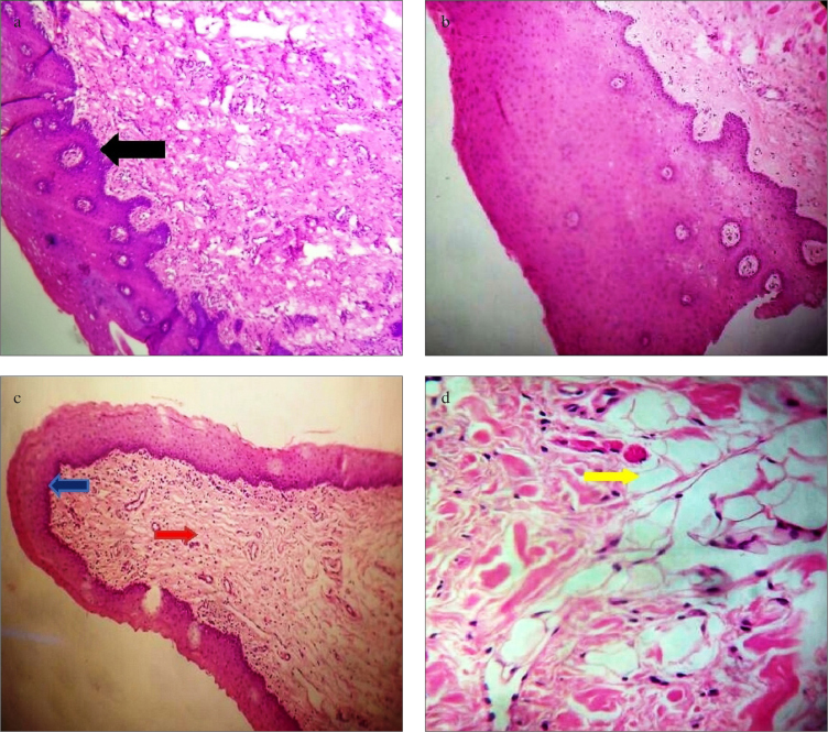
Figure 2.
a–d. Histopathological characteristics of lingual mucosa. (a, b): Graft harvested from ventral surface of tongue showing stratified squamous epithelium (black arrow) (10× and 40× magnification). (c): Integrated lingual mucosal graft showing lymphocytic infiltration (red arrow) with maintained epithelial characteristics (blue arrow) (10× magnification). (d): Under 40 magnification, these integrated graft showing vacuolar degenerations (yellow arrow)

