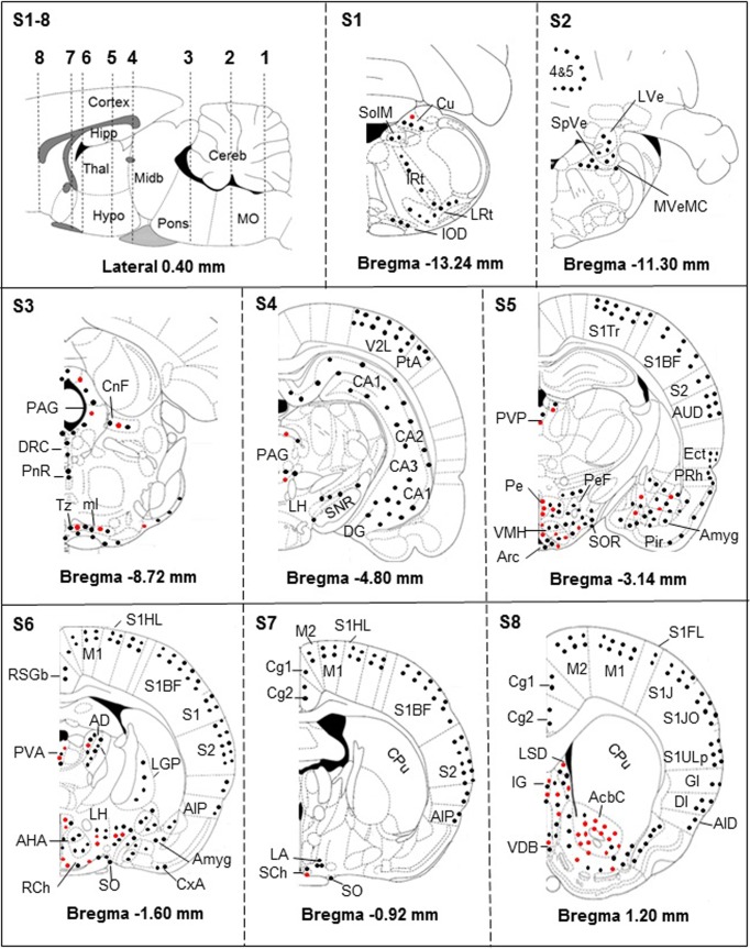
Fig 2. Brain distribution of iNOS-positive neurons (black dots), astrocytes and microglial cells (red dots).
S1-8 –Sagittal schema of the brain (lateral 0.4 mm; according to [25]) showing the 8 sections (S1-S8) performed in 4 rats. The sections extended from bregma +1.20 mm to bregma -13.24 mm and were analyzed for the distributions of the cells. The iNOS-positive astrocytes and microglial cells are mainly located in periventricular structures and basal parts of the brain. The cerebral cortex remains devoid of iNOS-positive astrocytes and microglial cells (S4, S5, S6, S7 and S8). Abbreviations: n, nucleus; 4&5, Cerebellar lobules; AcbC, accumbens n, core; AHA, anterior hypothalamic area, anterior part; AID, agranular insular cortex, dorsal; AIP, agranular insular cortex, posterior; Amyg, amygdala; Arc, arcuate n; AUD, secondary auditory cortex, dorsal; CA1, CA2, CA3, hippocampus fields (corni ammonis CA1, CA2 and CA3); Cortex, cerebral cortex; Cereb, cerebellum; Cg1, cingulate cortex, area 1; Cg2, cingulate cortex, area 2; CnF, cuneiform n; CPu, caudate putamen; Cu, cuneate n; CxA, cortex amygdala transition zone; DG, dentate gyrus; DI, dysgranular insular cortex; DRC, dorsal raphe n, caudal part; Ect, ectorhinal cortex; GI, granular insular cortex; Hipp, hippocampus; Hypo, hypothalamus; IG, granular insular cortex; IOD, inferior olive, dorsal n; IRt, intermediate reticular n; LA, lateroanterior hypothalamic n; LGP, lateral globus pallidus; LH, lateral hypothalamic area; LRt, lateral reticular n; LSD, lateral septal n, dorsal part; LVe, lateral vestibular n; M1, primary motor cortex; M2, secondary motor cortex; Midb, midbrain; ml, medial lemniscus; ml, Medial Lemniscus; MO, medulla oblongata; MVeMC, medial vestibular n, magnocellular part; PAG, periaqueductal gray; Pe, periventricular hypothalamic n; PeF, perifornical n; Pir, piriform cortex; PnR, pontine raphe n; PRh, perirhinal cortex; PtA, parietal association cortex; PVA, paraventricular hypothalamic n, anterior part; PVP, paraventricular thalamic n, posterior part; RCh, retrochiasmatic area; RSGb, retrospenial granular b cortex; S1, primary somatosensory cortex; S1BF, primary somatosensory cortex, barrel field; S1FL, primary somatosensory cortex, forelimb region; S1HL, Primary somatosensory cortex, hindlimb region; S1J, primary somatosensory cortex, jaw region; S1JO, primary somatosensory cortex, jaw region; S1Tr, primary somatosensory cortex, trunk region; S1ULp, primary somatosensory cortex, upper lip region; S2, secondary somatosensory cortex; SCh, suprachiasmatic n; SHi, septohippocampal n; SNR, substantia nigra; SO, supraoptic n; SolM, solitary n medial tract; SOR, supraoptic n, retrochiasmatic; SpVe, spinal vestibular n; Thal, thalamus; Tz, trapezoid body n; V2L, secondary visual cortex, lateral area; VDB, n of vertical limb diagonal band; VMH, Ventromedial thalamic. For a more detailed description of the structures, see [25].

