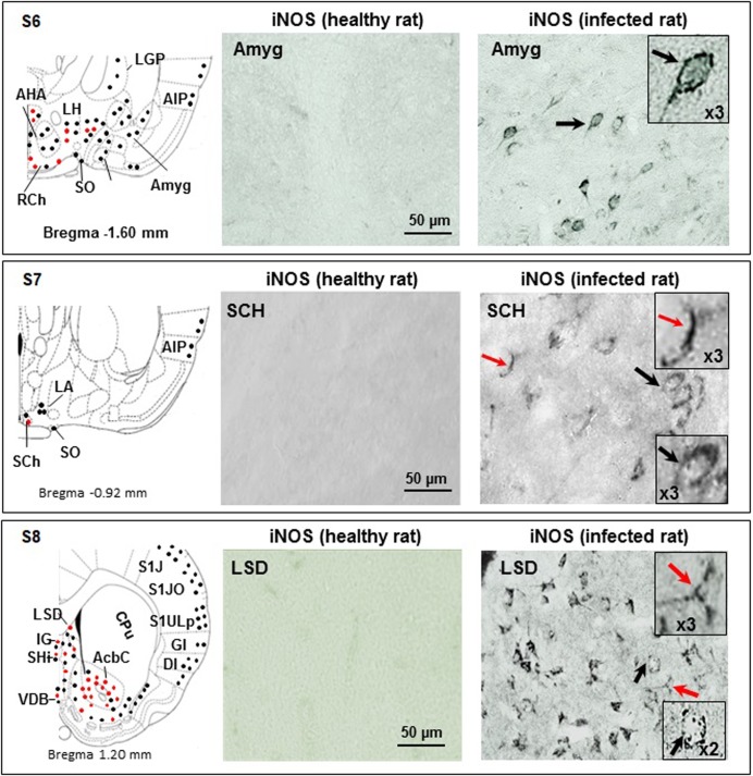
Fig 5. Immunohistochemical iNOS labelling observed in the coronal brain slices obtained through sections S6, S7 and S8.
Section S6 (bregma -1.60 mm): in control rats, Amyg is free of iNOS-positive cells; infected rats exhibit Amyg iNOS-positive neurons (black arrows and dots, also in X3 magnification); areas close to the 3rd cavity (AHA, LH, RCh and SO) contain iNOS positive neurons and neuroglial cells (black and red dots); the LGP exhibits only iNOS positive neurons (black dots); the amygdalian cortex (CxA) and the agranular insular cortex (AIP) contain only iNOS positive neurons (black dots). Section S7 (bregma -0.92 mm): in control rats, the suprachiasmatic nuclei (SCh) do not reveal iNOS-positive cells; in infected rats, SCh exhibit iNOS-positive astrocytes (red arrow and dots) and neurons (black arrow and dots); magnifications (X3) are also shown; in LA and more superficial structure (AIP), only iNOS positive neurons are observed (black dots in schema). Section S8 (bregma 1.20 mm): in control rats, the LSD does not reveal iNOS-positive cells, contrary to infected rats, who exhibit iNOS-positive astrocytes and microglial cells (red arrows and dots) and neurons (black arrows and dots). Magnifications (X3) are also shown; in the ventral part of the slice, iNOS positive neuroglial cells are abundant in AcbC and in a lesser extent in IG and VDB (red and black dots); again, superficial structures (AID, DI, GI, S1J, S1JO and S1ULp) exhibit only iNOS positive neurons (black dots). The CPu does not exhibit any iNOS positive cell. Abbreviations and signs see Fig 2 and reference [25].

