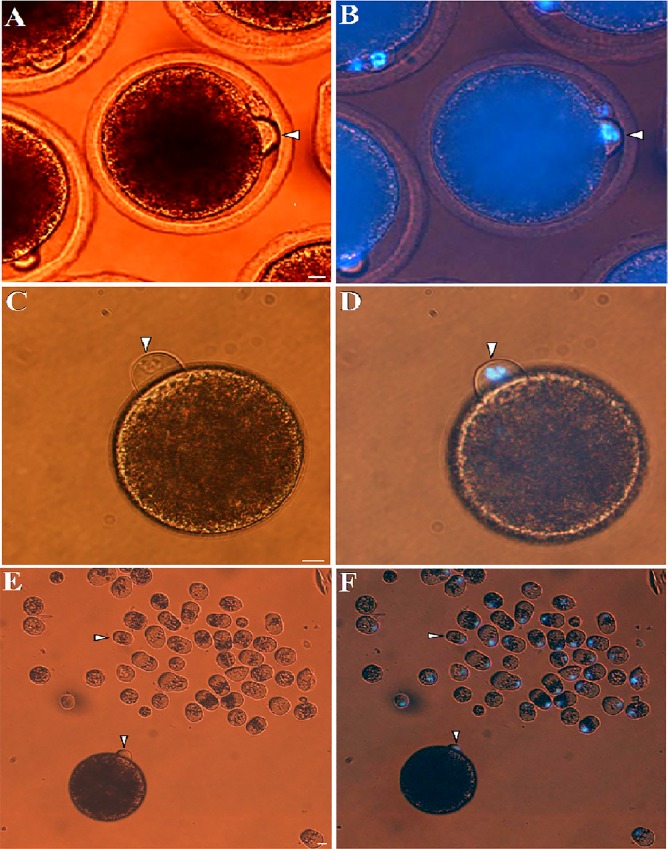
Fig 2. Cytoplasmic protrusion of MII-spindle for m-HMC in dromedary camel.
In vitro matured dromedary camel oocytes with characteristic cytoplasmic protrusion (arrow head) observed under bright light (A) and UV-light (B). A typical protrusion (white arrow head) of matured oocytes treated after zona removal and demecolcine treatment observed under bright light (C) and UV-light (D). MII cytoplasmic protrusions (arrow head) removed from MII oocytes using m-HMC method compared with an intact oocyte (asterisk) under bright light (E) and UV-light (F). Bar = 10 μm.

