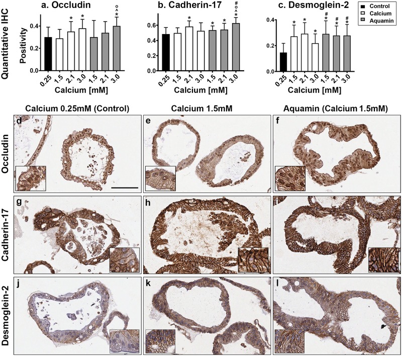
Fig 5. Cell surface components.
At the end of the incubation period, colonoids were examined after immunostaining of histological sections. Quantitative immunohistochemical analysis (a-c) and images: occludin (d, e and f); Cadherin-17 (g, h and i); desmoglein-2 (j, k and l). Quantitative IHC. Positivity measured using Positive Pixel Value algorithm. Means and SDs based on 26 to 100 individual crypts in each condition. Asterisks (*) indicate statistical significance from control. Additional symbols indicate statistical significance as follows: ^ from calcium 1.5mM, O from calcium 2.1mM, and # from calcium 3.0mM, with a significance at p<0.05 level. All three proteins are visible in colonoids under all conditions by immunohistology. With desmoglein-2, there is a clear shift from cytoplasmic to surface with intervention. Bar for (d-l) main panel = 100μm. Insets: respective stained colonoids at a higher resolution.

