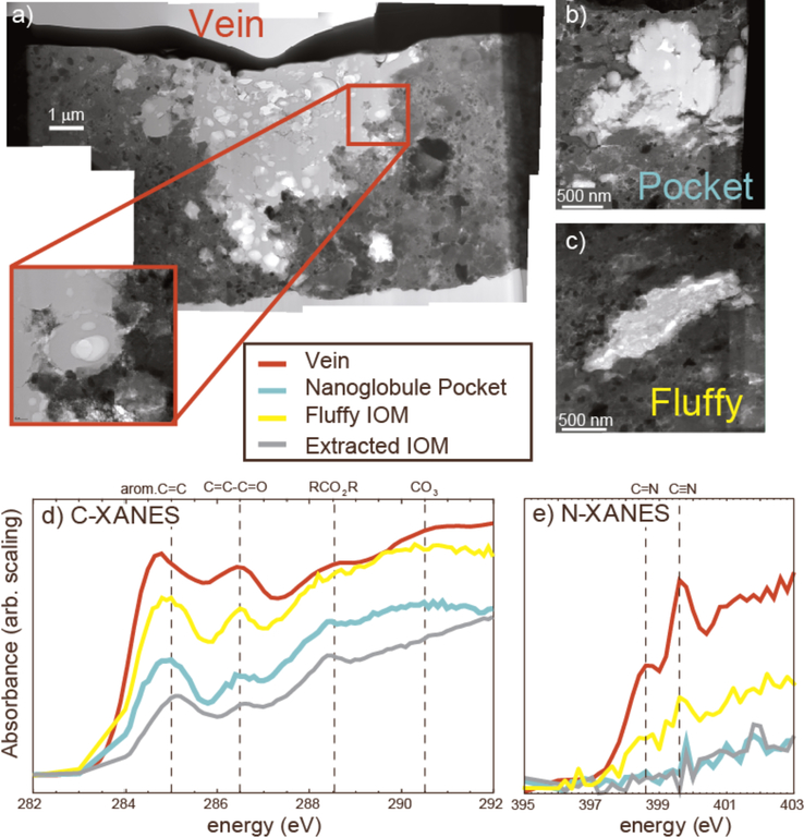
Figure 3.
Coordinated in situ microanalyses of organic matter in QUE 99177 (CR2). (a) A bright field STEM image mosaic of a FIB section cut through the organic-rich vein in Figure 2, which appears to be an aggregate of nanoglobules (see inset). Figures (b) and (c) are bright field STEM images of organic inclusions in another QUE 99177 FIB section, a small aggregate (“pocket”) of nanoglobules and a carbonaceous particle with a fluffy texture, respectively. Figures d) and e) are C-XANES and N-XANES spectra, respectively, of organic features indicated in the STEM images compared to the average spectra of IOM extracted from the same meteorite. The XANES measurements reveal heterogeneity in functional-group chemistry on a μm scale. There is a much stronger nitrile peak associated with this vein than in the fluffy IOM and bulk extracted IOM.

