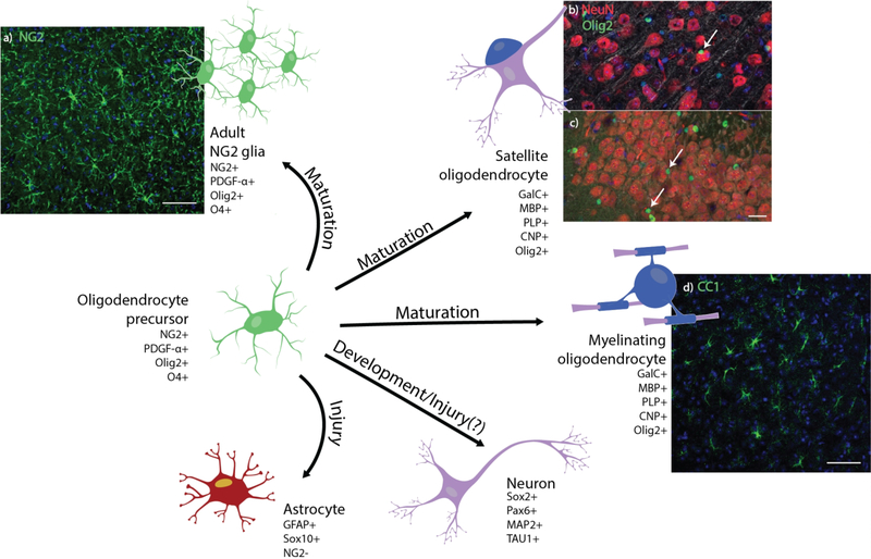
Figure 1. Development and cell fate of oligodendrocyte precursor cells.
Oligodendrocyte precursor cells maintain various paths of cell fate depending on tissue conditions. (a) Histological stain of NG2+ glia (green) and cell nuclei (blue) in horizontal sections of the adult mouse cortex. Scale bar = 100 μm. Olig2+ oligodendrocytes (green) can be perineuronal (white arrow), residing in close proximity to NeuN+ neuron cell bodies (red) in both the (b) cortex and (c) dentate gyrus of coronal sections of the adult mouse brain. Scale bar = 25 μm. (d) Confocal image of cortical CC1+ myelinating oligodendrocytes (green) and cell nuclei (blue) in horizontal sections of the adult mouse. Scale bar = 100 μm. Oligodendrocyte precursors have been observed in certain regions of the brain to transdifferentiate into neurons and, under specific injury conditions, into reactive astrocytes.

