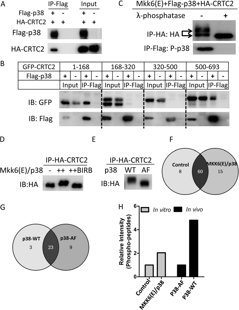
FIG 5.
CRTC2 is hyperphosphorylated by p38. (A) Western blot of coimmunoprecipitation of Flag-p38 with HA-tagged-CRTC2 (HA-CRTC2) in HEK293T cells. Coimmunoprecipitation (co-IP) was performed using anti-Flag M2 beads. Antibodies against Flag and HA were used for Western blotting. (B) Western blot of coimmunoprecipitation of Flag-p38 with GFP-tagged-CRTC2 truncates (CRTC2 aa 1 to 168, 168 to 320, 320 to 500, and 500 to 693) in HEK293T cells. Coimmunoprecipitation was performed using anti-Flag M2 beads. Antibodies against Flag and HA were used for Western blotting. (C) In vitro dephosphorylation assay of MKK6(E)/p38-phosphorylated CRTC2. HA-CRTC2 was purified from cells coexpressing MKK6(E)/Flag-p38 using anti-HA beads and incubated with λ-phosphatase. Purified Flag-p38 was used as a positive control. Antibodies against Flag and HA were used for Western blotting. (D) In vitro kinase assay for HA-CRTC2. HA-CRTC2 purified from overexpression in HEK293T cells was incubated with recombinant MKK6(E) and p38 in the absence of the p38 inhibitor BIRB-796 (10 μM). Antibodies against Flag and HA were used for Western blotting. (E) In vivo phosphorylation of HA-CRTC2 in HEK293T cells. Cells were cotransfected with HA-CRTC2 and MKK6(E)/p38-WT or MKK6(E)/p38-AF. HA-CRTC2 was purified from cells coexpressing MKK6(E)/Flag-p38 using anti-HA beads. An antibody against HA was used for Western blotting. (F) Venn diagram of the phosphorylation sites of CRTC2 identified by LC-MS/MS assay from the experiment shown in panel D. Detailed sites are shown in Table 1. (G) Venn diagram of the phosphorylation sites of CRTC2 identified by LC-MS/MS assay from the experiment shown in panel E. Detailed sites are shown in Table 2. (H) Quantification of the intensity of phosphopeptides obtained for panels F and G.

