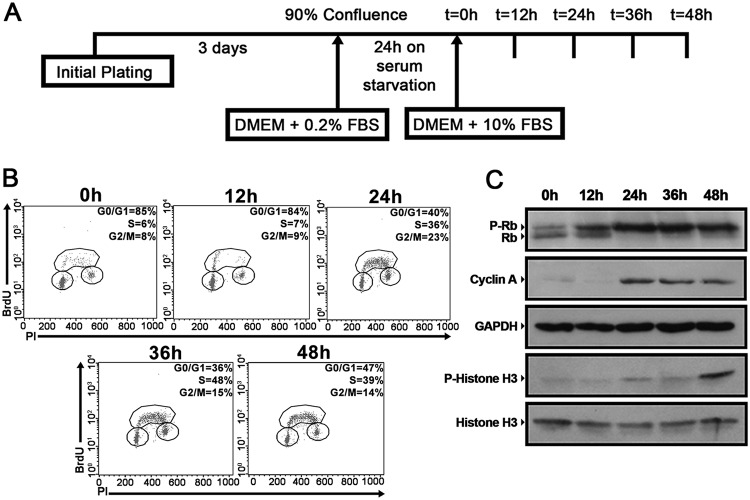
FIG 1.
Synchronized NIH 3T3 cells progress uniformly through the cell cycle upon serum supplementation. (A) Experimental scheme of NIH 3T3 cell synchronization. NIH 3T3 cells were cultured for 72 h until 90% confluence and were then maintained on serum starvation for 24 h. The cells were subsequently replated at a low density, supplemented with a medium containing 10% FBS, and analyzed at the indicated time points. (B) Synchronized NIH 3T3 cells were incubated with 20 μM BrdU for 30 min before fixation at the indicated time points. Later, cell cycle analysis was performed using FITC-conjugated anti-BrdU antibodies together with propidium iodide staining. The percentage of cells in each stage of the cell cycle (G0/G1, S, and G2/M) is indicated. (C) Whole-cell extracts were prepared from synchronized NIH 3T3 cells at the indicated time points and were separated by SDS-PAGE. Western blotting was performed using antibodies to the indicated proteins. All data are representative of the results of at least three independent experiments.

