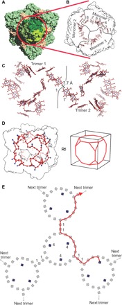
Fig. 2. Electron transfer network in HDH.

(A) Entire HDH assembly, with one trimer (front, red circle) shown in different shades of green. (B) Heme network in a trimer of HDH. The eight heme groups of each monomer form a ring-like relay system for electrons, connecting the active site heme 4 moieties to the exit sites for electrons at heme 1 as in a typical HAO-like enzyme. (C) Heme networks of two individual trimers in the HDH complex. Heme 1 of the one trimer is in close proximity to a heme 1 of the other trimer, likely allowing efficient electron transfer. Their edge-to-edge distance is indicated. (D) Proposed network of heme groups in the HDH complex. Each heme group is represented by its iron atom, shown as a red sphere or a blue sphere in case of an active site heme 4 iron. The surface of the HDH complex is shown as a black outline. The heme network approximates a truncated cube. (E). Schematic of part of the heme network in the HDH complex. Active site hemes (labeled “4”) are shown in solid blue, and other heme groups are shown as open black symbols. A possible path for electrons from one active site to a distant trimer in the complex is indicated by the red line.
