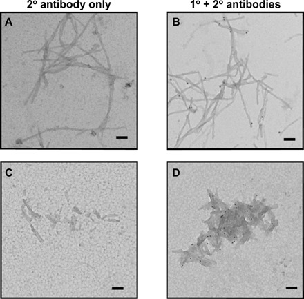
Fig. 2. Analysis of clusterin interactions with Aβ(M1-42) fibrils using immunogold TEM.

Aβ(M1-42) fibrils formed under quiescent condition imaged as is (A and B) and after sonication (C and D) were incubated with BSA and clusterin and stringently washed. Incubation with an anti-mouse secondary antibody conjugated to a gold particle showed no nonspecific labeling (A and C), whereas incubation with an anti-clusterin monoclonal antibody followed by an anti-mouse secondary antibody conjugated to a gold particle shows the presence of clusterin interacting with the Aβ(M1-42) fibrils (black dots). Scale bars, 100 nm.
