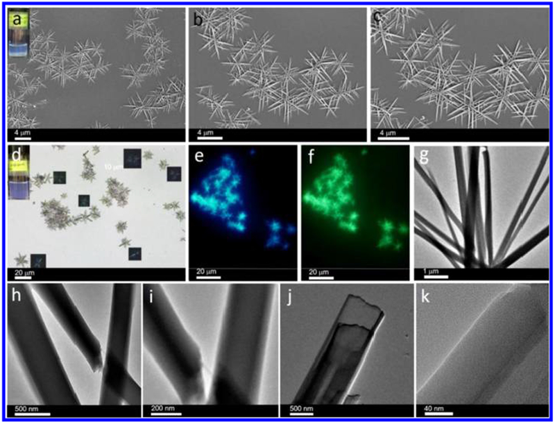
Figure 1.

(a) SEM images of microneedle-based microflowers formed by cage 1 (10 μM) in a DCM/EA mixture with 80% EA (inset: optical image showing the assembly method in binary solvents). (b,c) Corresponding assemblies at different magnifications. (d) Optical microscopy image of cage 1-based assemblies (10 μM) over a large area (inset: corresponding digital photo and polarized optical microscopy image). (e,f) Fluorescence microscopy images of these assemblies under (e) ultraviolet-light excitation and (f) blue-light excitation. (g) TEM image of microflowers formed by cage 1 (10 μM). (h) TEM image showing the intersection between two needles. (i,j) TEM images showing the multilayer structures. (k) Magnified TEM image of the layer structure.
