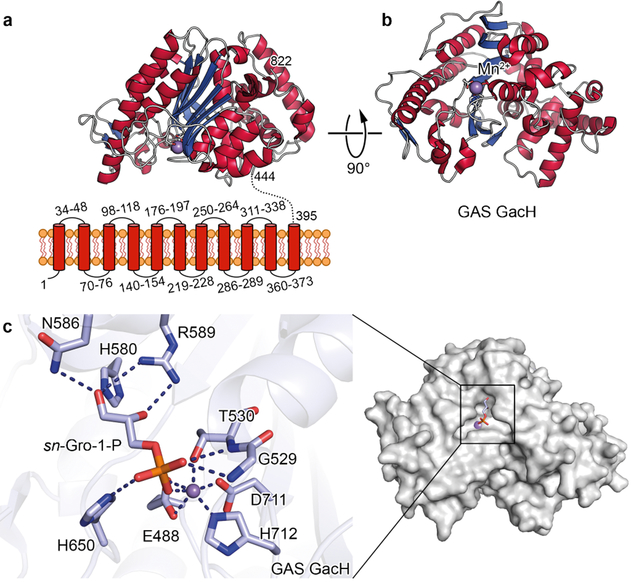
Fig. 3. Structure of eGacH.
a, Predicted topology of GacH showing eleven transmembrane helices and structure of extracellular domain with the enzymatic active site oriented towards the cell membrane. b, Structure of apo eGacH viewing at the active site with the Mn2+ ion shown as a violet sphere. c, A close-up view of the active site GacH crystal structure in complex with sn-Gro-1-P.

