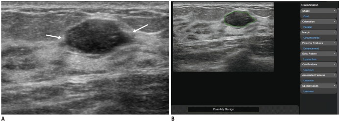
Fig. 1. 24-year-old woman diagnosed with fibroadenoma using US-guided biopsy.
A. Transverse B-mode US image shows 15-mm oval hypoechoic mass (arrows). B. After radiologist clicked on center point of mass on US image shown, two-dimensional region of interest (green line) was automatically drawn along mass margin through deep learning-based CAD. Following this, deep learning-based CAD analyzed US features of mass according to BI-RADS lexicon and displayed final assessment of “possibly benign” on screen. During first reading session (US images alone), two readers classified mass as BI-RADS category 4a because they assessed that margin of mass was angular (right arrow in A), whereas other two readers did not and classified mass as category 3. During second reading session (US images with CAD), two readers who previously classified mass as category 4a reassessed it as category 3, whereas two readers who previously classified it as category 3 did not change their classifications. BI-RADS = Breast Imaging Reporting and Data System, CAD = computer-aided diagnosis, US = ultrasound

