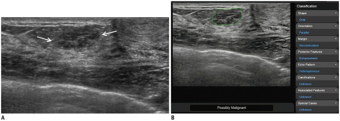
Fig. 3. 50-year-old woman diagnosed with ductal carcinoma in situ using US-guided biopsy and surgical excision.
A. Transverse B-mode US image shows 13-mm oval mass with slightly heterogeneous echo pattern (arrows). B. Deep learning-based CAD analyzed US features of mass (green line) and displayed final assessment of “possibly malignant” on screen. During first reading session (US images alone), all four readers classified mass as BI-RADS category 3. During second reading session (US images with CAD), three of four readers changed their assessment to category 4a.

