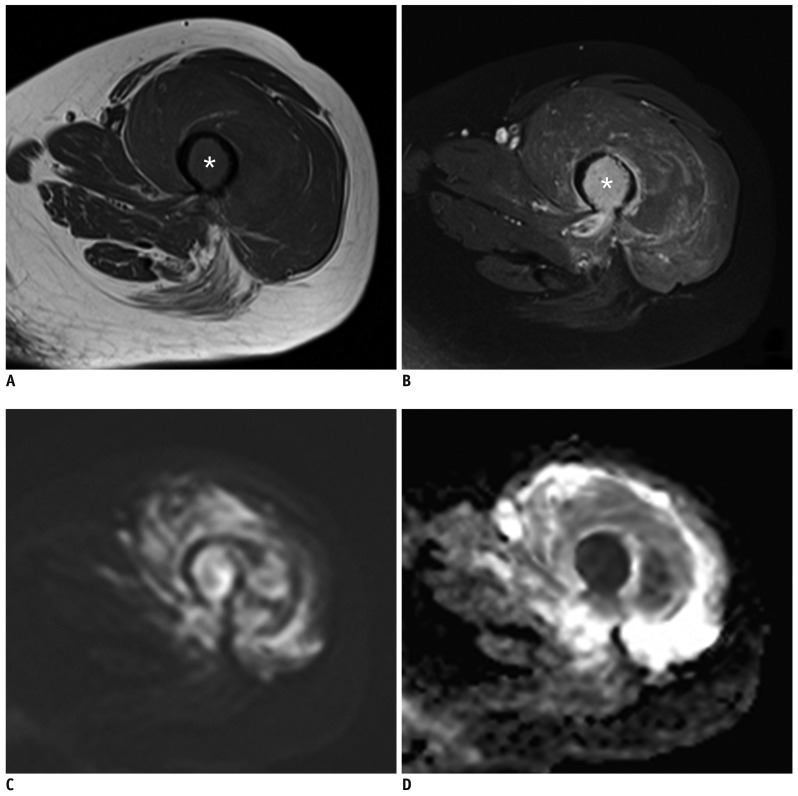
Fig. 2. CE and combined sets demonstrate correct diagnosis of pathologic fracture by both readers at left femur in 55-year-old woman with multiple myeloma.
(A) Axial T1-weighted, and (B) axial contrast-enhanced fat-saturated T1-weighted images show intramedullary enhancing single bone abnormality at fracture site with perilesional edema (asterisks). (C) Corresponding axial DWI shows high signal change at fracture site (b = 1400 s/mm2). (D) Axial ADC map shows impeded water diffusivity. ADC = apparent diffusion coefficients

