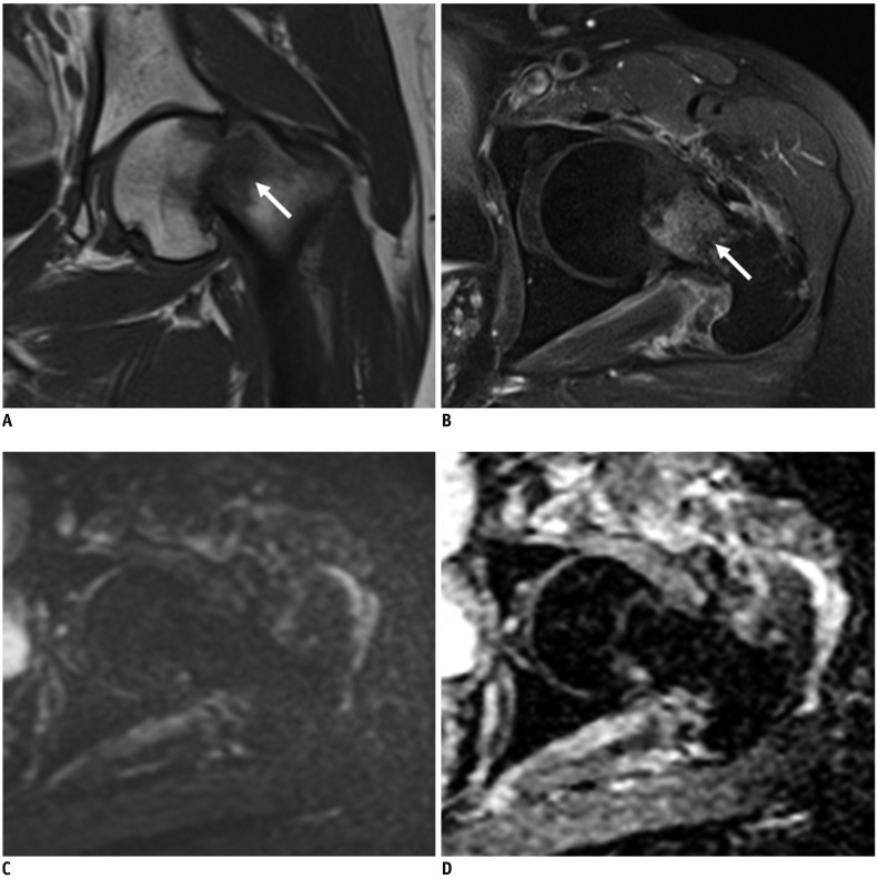
Fig. 3. CE and combined sets demonstrate discordant interpretations of traumatic fracture at left femur in 56-year-old woman with vaginal cancer.
Initial diagnosis of reader 1 from CE set was pathologic fracture due to focal bone abnormality on T1-weighted images with enhancement at fracture margin, in which DWI is added to CE set, reader 1 changed diagnosis to traumatic fracture. (A) Coronal T1-weighted and (B) axial contrast enhanced fat-saturated T1-weighted images show single bone abnormality with bone marrow enhancement at fracture site in left proximal femur, neck (arrows). (C) Corresponding axial DWI shows no high signal at fracture site (b = 1400 s/mm2). (D) Axial ADC map shows no impeded water diffusivity. This case was pathologically confirmed as intramedullary hemorrhage with traumatic fracture.

