. 2019 Jan 16;43(2):192–205. doi: 10.4093/dmj.2018.0052

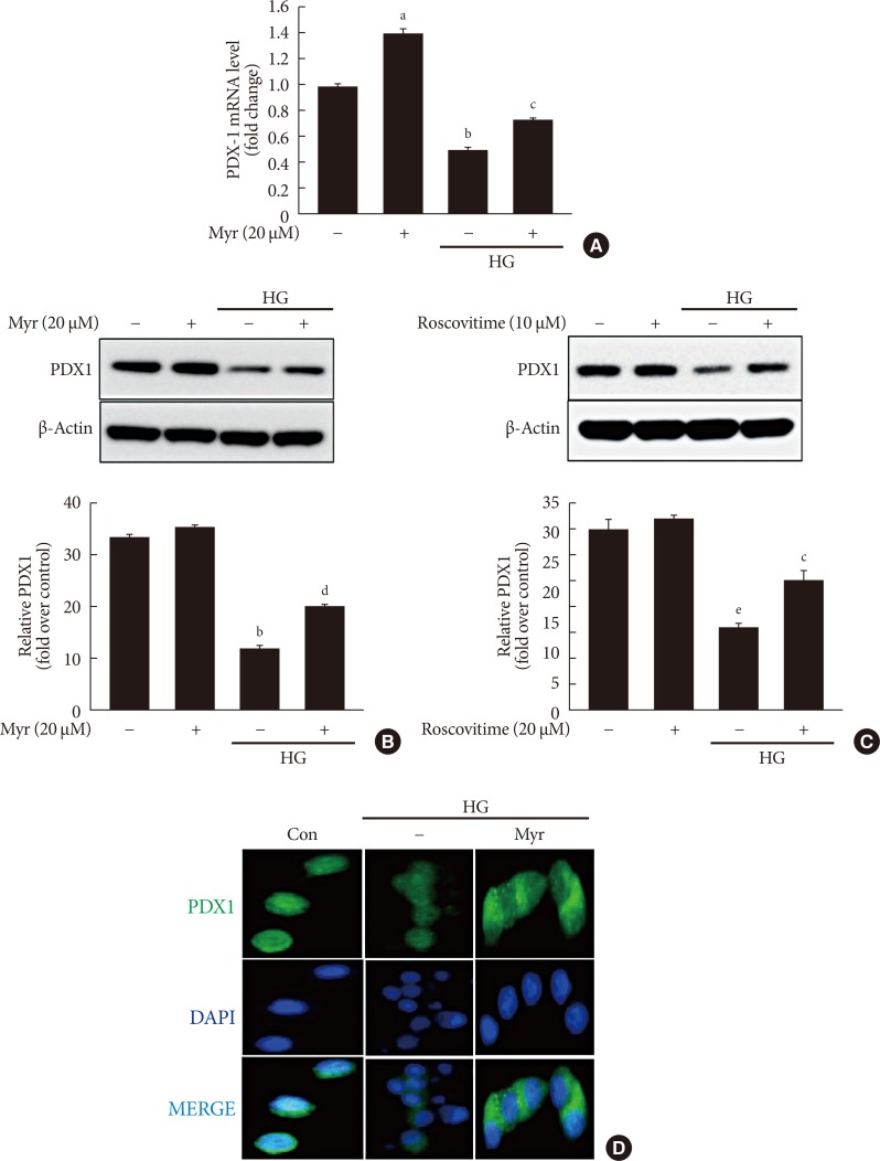
Fig. 4. Myricetin counteracts the decrease in total and nuclear levels of pancreatic duodenal homeobox 1 (PDX1) in high glucose (HG)-exposed INS-1 cells. (A, B) INS-1 cells were incubated with 30 mM glucose (HG) in the presence or absence of myricetin for 24 hours. (A) PDX1 mRNA levels were determined by real-time polymerase chain reaction (PCR). Data are expressed as the mean±standard deviation of at least three independent experiments. (B) Representative images of Western blot analysis of PDX1. (C) INS-1 cells were incubated with 30 mM glucose (HG) in the presence or absence of roscovitine for 24 hours. Representative images of western blot analysis of PDX1. (D) INS-1 cells were incubated with 30 mM glucose (HG) along with myricetin for 24 hours. Representative images of the subcellular localization of PDX1 by confocal microscope. 4′,6-Ddiamidino-2-phenylindole (DAPI) was used to stain the nuclei. aP<0.05 vs. control, bP<0.001 vs. control, cP<0.05 vs. HG, dP<0.01 vs. HG, eP<0.01 vs. control.

Fig. 4