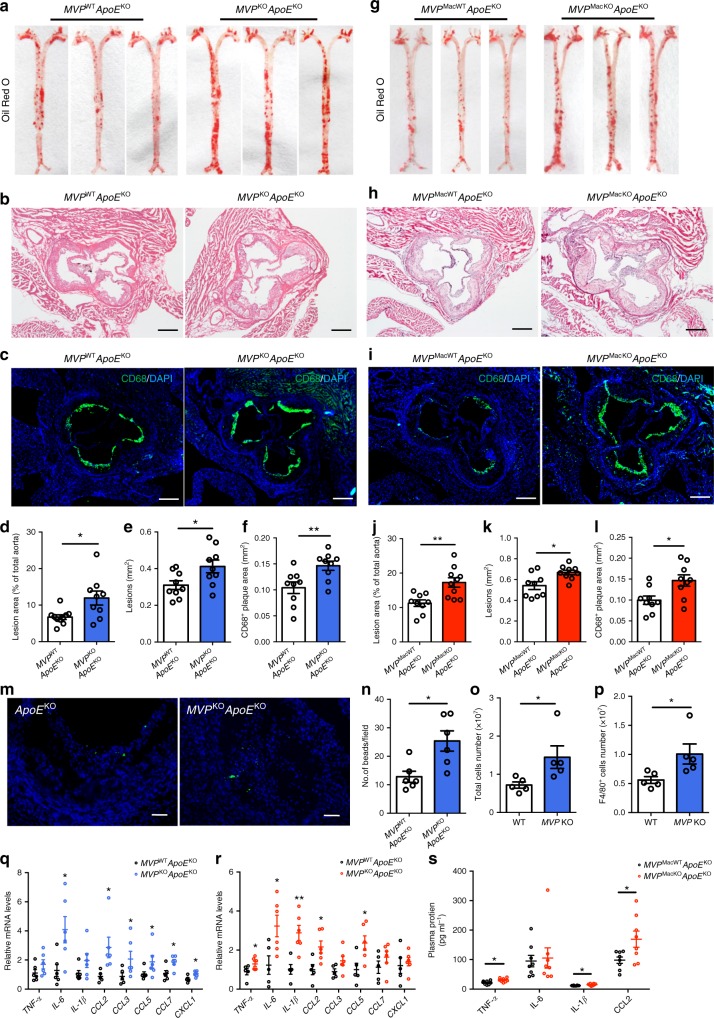
Fig. 5.
Deficiency of MVP accelerates atherosclerosis progression. a, d En face Oil Red O staining of whole aortas from MVPKOApoEKO (n = 9) and control MVPWTApoEKO (n = 10) male mice fed with a WD for 10 weeks (a). Lesion occupation was quantified and shown in (d). b, e Representative H&E-stained images (b) and quantitative analysis (e) of the lesions in aortic root sections from MVPKOApoEKO and MVPWTApoEKO mice (n = 9). Quantification of lesion burden was performed by cross-sectional analysis of the aortic root. Scale bars, 200 μm. c, f Representative CD68+ staining in cross-sections (c) and quantitative analysis (f) of the aortic root plaques from MVPKOApoEKO and MVPWTApoEKO mice (n = 9). Scale bars, 200 μm. g, j En face Oil Red O staining of aortas from MVPMacKOApoEKO (n = 10) and control MVPMacWTApoEKO (n = 9) mice fed a WD for 12 weeks (g). Lesion occupation was quantified and shown in (j). h, k Representative H&E-stained images (h) and quantitative analysis (k) of the lesions in aortic root sections from MVPMacKOApoEKO and MVPMacWTApoEKO mice (n = 9). Scale bars, 200 μm. i, l Representative CD68+ staining in cross-sections (i) and quantitative analysis (l) of the aortic root plaques from MVPMacKOApoEKO and MVPMacWTApoEKO mice (n = 9). Scale bars, 200 μm. m, n Quantitative analysis of infiltrated fluorescent bead-labeled monocytes in atherosclerotic lesions of MVPKOApoEKO and MVPWTApoEKO mice fed with a WD for 10 weeks (n = 6). o, p Three days after intraperitoneal injection of 1 ml 4% sterile thioglycollate media, total number of peritoneal cells (o) and F4/80+ PMs (p) of WT and MVP KO mice were measured (n = 5). q, r mRNA levels of inflammatory mediators in the aortas of MVPKOApoEKO (q) and MVPMacKOApoEKO (r) mice (n = 5–6). s Plasma concentration of TNF-α, IL-6, IL-1β, and CCL2 in MVPMacKOApoEKO and MVPMacWTApoEKO mice (n = 8). Data are expressed as mean ± SEM. *P < 0.05 and **P < 0.01 by Student’s t test

