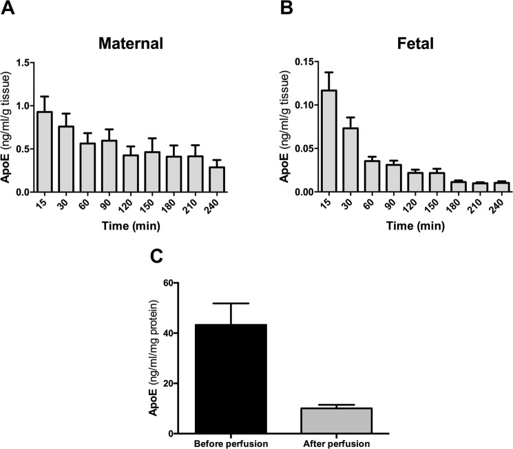
Figure 2.
Secretion of apoE in the human perfused placenta. Four term human placentas were initially perfused for 30 minutes with DMEM medium and EBSS buffer containing 2 g/L of glucose, 10 g/L Dextran FP40, 40 g/L BSA and 2.5 IE/mL heparin. This washing phase was followed by 240 minutes perfusion of the maternal and fetal circulation as an open/open system. Samples were taken at 15, 30, 90, 120, 150, 180, 210 and 240 minutes. (A,B) The concentration of apoE in the maternal (A) and the fetal perfusate (B) was measured at each time point by ELISA and normalized to the weight of the cotyledon (g). (C) Measurement of apoE concentrations in placental tissue lysates before and after the perfusion by ELISA. The concentration was normalized to the total protein content (mg). Results represent mean ± SEM of four independent experiments measured in triplicates; *p < 0.05.

