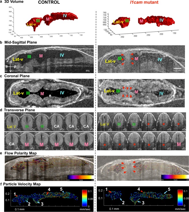
Figure 4.
F0 CRISPR mutation in L1CAM causes cerebral aqueduct stenosis. The left column shows control and right column shows l1cam F0 CRISPR mutant images for all panels. (a) 3D rendering of the tadpole ventricular system shows aqueductal stenosis and a smaller ventricular system in l1cam F0 CRISPR mutant. (b) Mid sagittal view and (c) Coronal view of control and l1cam F0 CRISPR mutant, the later showing stenosis of the cerebral aqueduct (red arrowheads). (d) Transverse view of the control and l1cam F0 CRISPR mutant, starting at the end of the lateral ventricle through the cerebral aqueduct and ending in the midbrain ventricle. Control embryo shows normal opening of the duct whereas the mutant shows complete blockage (red star). (e) Relatively normal ciliary flow fields in the control and mutant animals. (f) 2D Particle Velocity Map shows intact FFs 1-5. (Lat-V: lateral ventricle, III: 3rd ventricle, M: Midbrain ventricle, IV: 4th ventricle).

