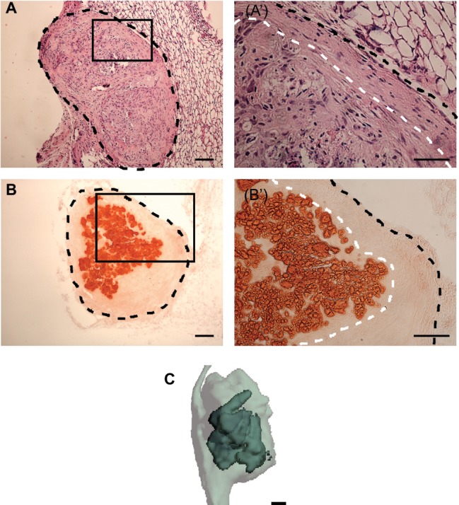
Figure 4.

H&E, alizarin red staining, and μCT analysis of scaffold-free PDLC constructs following in vivo implantation. (A) H&E staining of histological section of engineered tissue explant following 4-week subcutaneous implantations. Black, dashed line outlines explant. (Aʹ) Higher magnification image of boxed region in (A) shows the formation of two morphologically distinct tissues on the explant, a core tissue and a separate tissue on the periphery; black, dashed line outlines the sample and white, dashed line separates the inner and outer tissue structures. (B) Alizarin red staining of full explant following 4-week implantation. Black, dashed line outlines full explant. (Bʹ) Higher magnification image of boxed region in (B) shows that the core of the explant is stained positively for alizarin red while the periphery does not. Black, dashed line outlines the explant and the white, dashed line delineates the mineralized core tissue from the peripheral unmineralized tissue. (C) Three-dimensional rendering of μCT scan of explant shows mineralized tissue (dark, solid gray) localized within soft tissue (light, translucent gray). Scale bars: (A,B,Bʹ,C) = 100 μm, (Aʹ) = 50 μm.
