Figure 2.
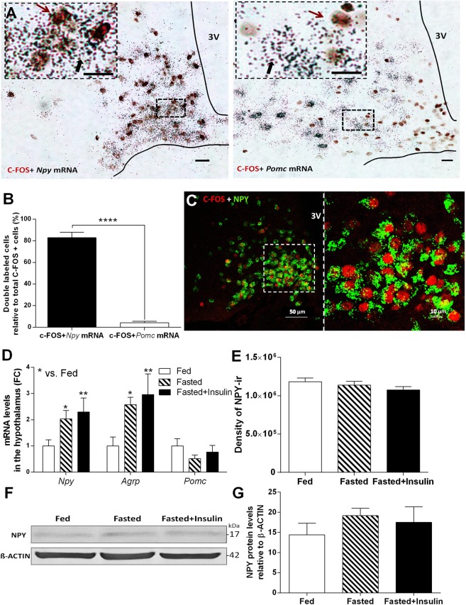
Hypoglycemia selectively activates orexigenic, NPY/AGRP -expressing neurons in hypothalamic arcuate nucleus (ARC). (A) Brightfield images of combined immunohistochemical (c-FOS; brown cell nuclei) and in situ hybridization histochemical [Npy or Pomc mRNA (black autoradiographic grains)] ARC preparations of fasted mice 1 h after insulin injection. Scale bars, 10 µm. (B) Colocalization of Npy or Pomc mRNA with c-FOS protein in ARC after insulin-induced hypoglycemia (n = 6–7). (C) Representative images of c-FOS (red) and NPY protein (green) colocalization in ARC of NPY-Ires-Cre ZsGreen reporter mice following insulin-induced hypoglycemia. (D) Expression of Npy, Agrp and Pomc mRNA in arcuate nucleus of fasted mice 1 h after insulin injection compared to fed and fasted, saline injected controls (n = 4 per groups). Fold change (FC) of mRNA expression was assessed by quantitative RT-PCR (qRT-PCR). (E) Quantitative analysis of NPY-immunostained hypothalamic sections of C57BL/6 mice. Bar graph represents the density of NPY-immunreactivity (ir) in the unit area of arcuate nucleus (n = 3 per groups). (F,G) NPY levels, representative Western blot image (F) and quantification (G) from hypothalamus of fed, fasted and fasted + insulin injected mice (n = 3-3 per group). Full-length blot is presented in Supplementary Fig. 3. Fed: fed, saline-injected group; Fasted: O/N fasted, saline-injected group; Fasted + insulin: O/N fasted, insulin-injected group. All data expressed as mean ± SEM. *p < 0.05, **p < 0.01 vs. fed. 3 V: third ventricle.

