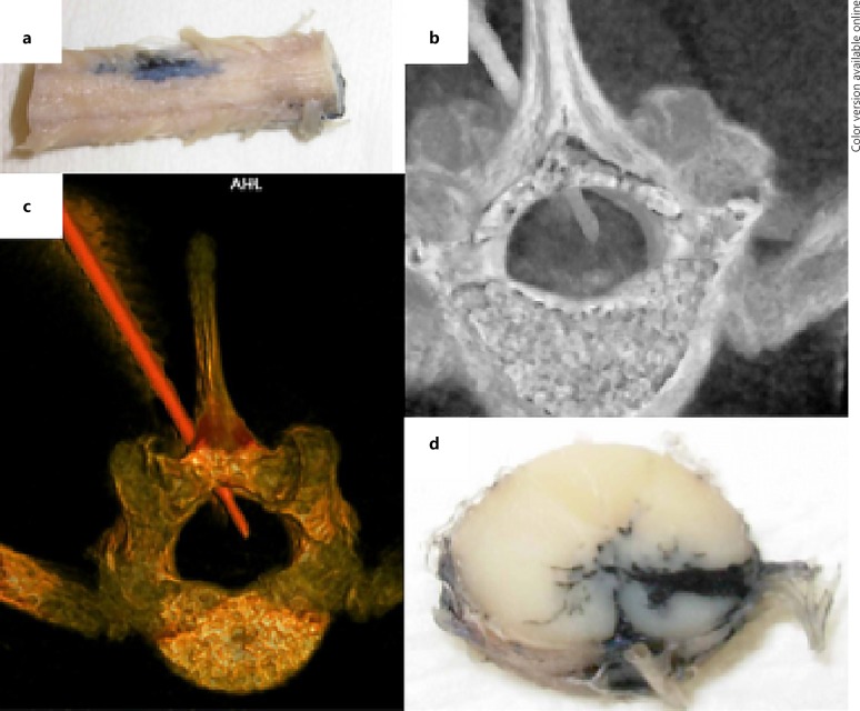
Fig. 4.
Studies of percutaneous delivery of India ink (to simulate drug delivery) into the spinal cord of a conventional swine. The figure shows representative images from past studies performed to explore spinal cord drug delivery approaches. a External anatomy of the infused spinal cord. b Computed tomography (CT) of the spine and spinal cord during delivery via percutaneous catheter. c Images of the vertebral bone and delivery catheter demonstrating the approach for subsequent. d Delivery into a targeted region within the ventral spinal cord.

