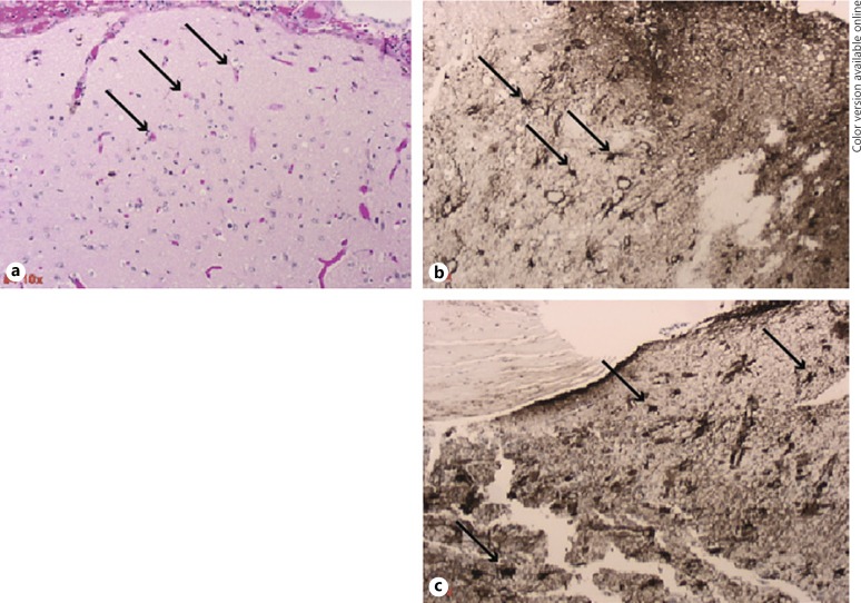
Fig. 3.
Gliosis associated with a closed loop neurostimulator device. H&E and GFAP stained sections of sub-temporal lobe cortical tissue attached to depth electrodes. Arrows show reactive astrocytes. a 10X H&E tissue section of the temporal lobe cortex previously attached to an anterior sub-temporal electrode. b, 10X GFAP stained tissue section as described in a. c, 10X GFAP section of temporal lobe cortex previously attached to a posterior sub-temporal electrode showing reactive astrocytes and gliosis.

