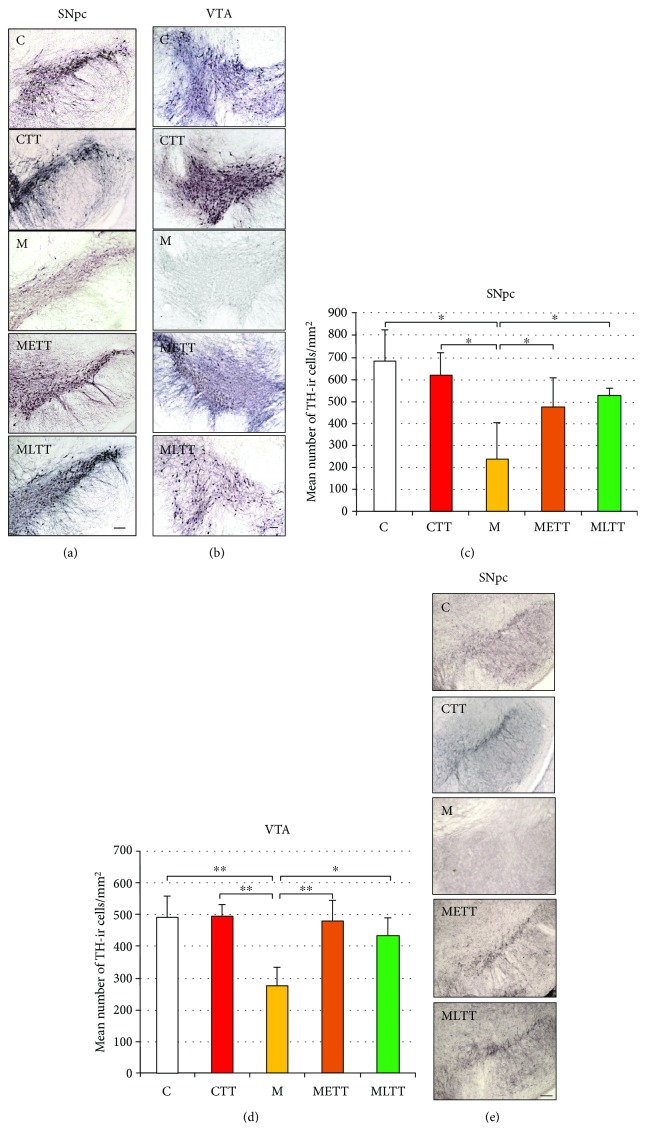
Figure 1.
Packing density of TH-immunoreactive neurons in the midbrain regions in the five experimental groups. Representative microphotographs of TH immunohistochemical staining in the substantia nigra pars compacta (SNpc) (a) and ventral tegmental area (VTA) (b), mean packing density of TH-immunoreactive neurons in the SNpc (c) and VTA (d), and microscopic images of VMAT2 immunohistochemical staining in the SNpc (e). C: control; CTT: control + treadmill training; M: treatment with MPTP; METT: MPTP treatment + early onset treadmill training; MLTT: MPTP treatment + late-onset treadmill training group. Scale bar: 100 μm. Statistical comparisons were performed with two-way ANOVA followed by Newman-Keuls post hoc test; ∗∗p < 0.001, ∗p < 0.05.

