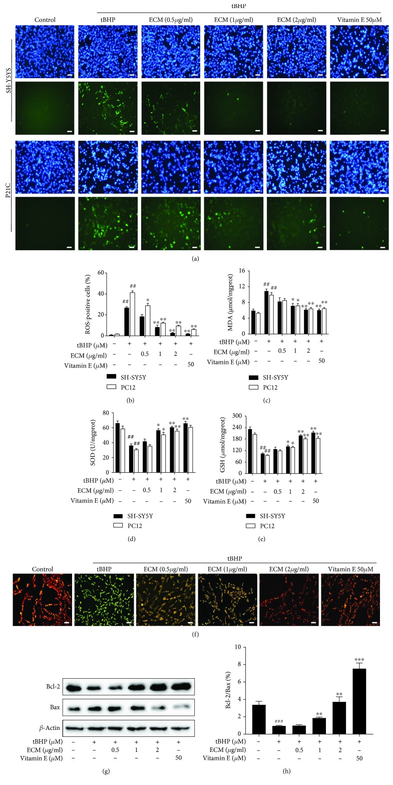
Figure 2.
ECM attenuates oxidative stress-induced mitochondrial dysfunction. The cells were pretreated with 0.5-2 μg/ml ECM and 50 μM vitamin E for 2 h and then exposed to 300 μM tBHP for an additional 6 h. (a) SH-SY5Y (upper panel) or PC12 cells (lower panel) were treated with DCFH-DA for 30 min; Hoechst 33342 was used to counterstain cell nuclei. Scale bar, 50 μm. (b) The percentage of ROS-positive cells among cultured SH-SY5Y or PC12 cells was quantified and was shown as histogram. Intracellular MDA content (c), SOD activity (d), and GSH levels (e) were detected using a kit assay and are presented as a histogram. (f) SH-SY5Y cells were pretreated with 0.5-2 μg/ml ECM for 2 h and then exposed to 300 μM tBHP for an additional 6 h. The mitochondrial membrane potential was determined using the JC-1 fluorescence probe, and representative pictures have been shown for comparison. (g) Western blot analysis was performed using antibodies against Bax and Bcl-2, and β-actin was used as a loading control. (h) The ratio of Bcl-2 to Bax was quantified by densitometry and is shown as a histogram. The results are shown as the mean ± SEM of three independent experiments. ## p < 0.01 in comparison with control cells. ∗ p < 0.05 and ∗∗ p < 0.01 in comparison with the cells exposed to tBHP alone.

