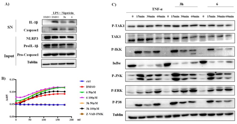
Figure 4.
(A) Immunoblot analysis of indicated proteins in culture supernatants (SN) and cell lysis (Input) of PMA primed THP-1 cells treated with target compounds (5 μM) and then stimulated with LPS (5 µg/mL) and NIG (20 µM). (B) Caspase 1 activity assay for purified caspase1 with or without presence of compounds 3h and 6 or positive control. (C) Immunoblot analysis of NF-κB and MAPK signaling pathway in 293 cells treated with TNF-α (20 ng/mL) with or without compounds 3h and 6 (5 μM). The protein levels were normalized against tublin.

