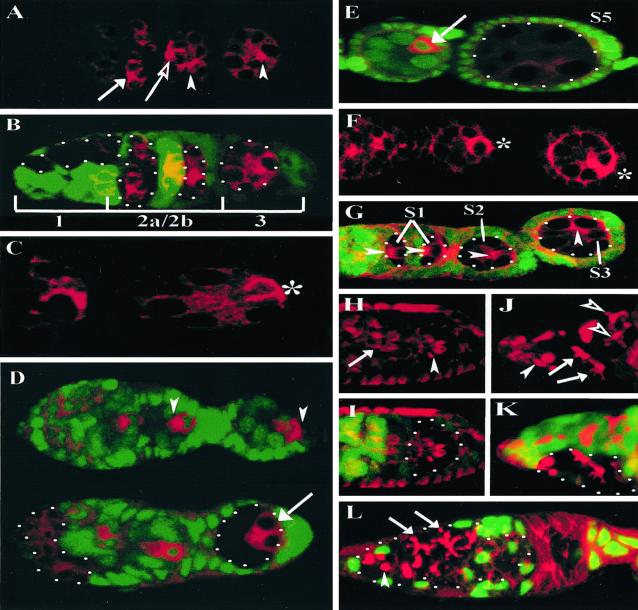
Figure 2.
BAZ and DaPKC are required to establish A-P polarity in the oocyte. (A and B) Germarium containing wild-type and baz null germ-line cysts double-labeled for ORB (red) and nuclear GFP (green) to mark baz clones (B, merge). (A) In region 2, both wild-type (open arrow) and baz null (arrowhead) germ-line cysts accumulate ORB in the two pro-oocytes. In contrast to wild-type (C, *), the baz null germ-line cyst in region 3 fails to translocate ORB from the anterior to a posterior crescent in the presumptive oocyte (A, arrowhead). (D) Wild-type and DaPKC mosaic germaria double-labeled for DHC64C (red) and nuclear GFP (green). In contrast to wild-type cysts (Upper) in which DHC64C localizes to a single posterior cell (arrowheads), in DaPKC mutant cysts (Lower) DHC64C fails to accumulate in a single cell but rather is localized to the two posterior-most cyst cells (arrow). (E) Wild-type and DaPKC mutant egg chambers double-labeled for DHC64C (red) and nuclear GFP (green) reveal that in contrast to wild-type (arrow), DaPKC mutant egg chambers fail to accumulate DHC64C. (F and G) In contrast to a wild-type germarium (F, *), analyses of baz null germ-line cysts up to stage 3 (S3) reveals a defect in the A-P MTOC transition (G, arrowheads). (H and I) Germarium double-labeled with rhodamine phalloidin (red) and nuclear GFP (green) reveals no defect in ring canal formation or spatial distribution within baz null germ-line cysts (H, arrowhead) as compared with a wild-type cyst (H, arrow). (J and K) Germarium double-labeled with 1B1 (J, red) and nuclear GFP (K, green, merge) indicates that baz mutant cysts display relatively normal fusome morphology albeit a somewhat weaker elaboration of fusome branches (J, open arrowhead), when compared with wild-type cysts (J, arrows). Spectrosome formation (J, filled arrowhead) is likewise unaffected in baz mutant germ-line clones. (L) DaPKC mosaic germarium double-labeled with 1B1 (red) and nuclear GFP (green) reveals no defect in spectrosome (arrowhead) or fusome assembly (arrows) in DaPKC mutant cysts. (Magnifications: A, B, D, and F–L, ×40; C, ×63; E, ×25.)

