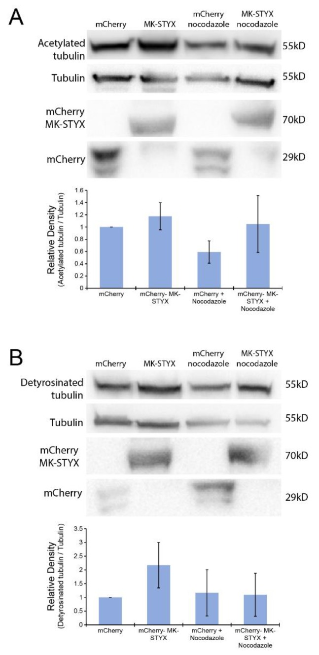
Figure 5.
MK-STYX increases acetylated and detyrosinated tubulin. Cells were transfected with expression plasmids for mCherry or mCherry-MK-STYX. Twenty-four hours post-transfection, cells treated with nocodazole or not were lysed and analyzed by immunoblotting. We examined cells expressing mCherry constructs by fluorescence microscopy to confirm transfection. (A) Blots were probed with anti-acetylated tubulin and showed that acetylated tubulin (55 kDa) is significantly decreased (paired t-test; p < 0.05) in the presence of nocodazole in control cells (mCherry-expressing) relative to cells expressing mCherry-MK-STYX. Cells overexpressing MK-STYX sustain acetylated tubulin in the presence of nocodazole. The blots were stripped and probed for tubulin as the loading control; they were also stripped and probed with anti-mCherry to confirm expression of mCherry (27 kDa) and mCherry-MK-STYX (67 kDa). (B) Lysates were also analyzed for detyrosinated tubulin (55 kDa) by detection with anti-detyrosinated tubulin. A significant increase (paired t-test; p < 0.05) in detyrosinated tubulin was observed in mCherry-MK-STYX-expressing cells in the absence of nocodazole, whereas detyrosination was decreased in cells expressing mCherry-MK-STYX in the presence of nocodazole. Blots were stripped and probed with anti-tubulin antibody for a loading control, then stripped a second time and probed with anti-mCherry antibody to confirm transfection. The error bars are ±SEM; three biologically independent replicate experiments were performed.

