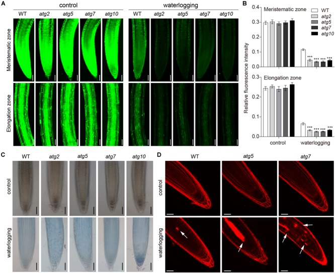
FIGURE 8.

Autophagy regulates the PCD process upon waterlogging. (A) Confocal images of the detection of cell viability in primary roots of atg mutants in comparison with wild type. Bars = 50 μm. (B) Meristematic and elongation zones of wild-type and atg mutants of FDA staining intensity are quantified by Image pro plus 6.0 software. (C) Trypan blue staining cell death in root of wild-type and atg mutants without treatment or waterlogged for 8 h. Bars = 50 μm. (D) Cell death of the root of wild-type and atg mutants visualized by PI staining. Arrows indicate the cell death. Bars = 50 μm. All of the experiments have been repeated at least three times. Data shown are the mean ± SD (n = 3). ∗P < 0.05; ∗∗P < 0.01; ∗∗∗P < 0.001 by Student’s t-test.
