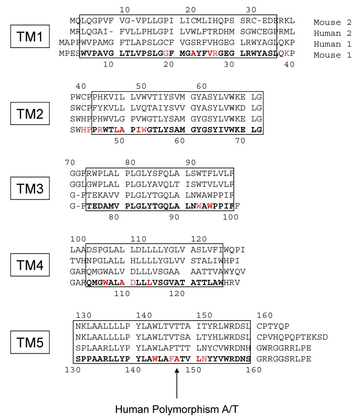
Figure 8.
The sequence alignment and helix assignment of isoforms 1 and 2 of mouse and human TSPO: The boxes depict the five transmembrane helices (labelled TM1 to TM5). The numbers on top correspond to the isoform 2 positions, whereas the numbers at the bottom correspond to isoform 1. The amino acids involved in the PK 11195 binding pocket of the atomic structure obtained from NMR data are written in red bold characters, and those observed at a short distance (3 angstroms) are in normal red characters.

