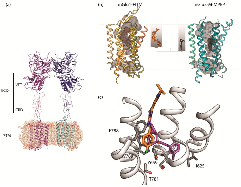
Figure 2.
The structure of mGlu receptors: (a) Structure of the full length apo mGlu5 receptor homodimer showing the VFT, CRD, and 7TM domains (PDB 6N52); (b) X-ray structures of mGlu1 and mGlu5 receptor 7TMs showing the different depth of binding for NAMs. (c) Close up view of the X-ray structures of the 7TM domains showing the overlay of NAMs FITM (orange) and mavoglurant (magenta) bound at mGlu1 and mGlu5 receptors with several amino acids labelled.

