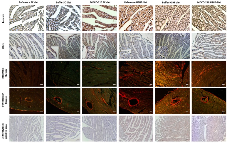
Figure 6.
Immunohistochemical and histochemical analysis of the myocardium of reference, buffer, and MDCO-216 SC diet and HSHF diet mice. Representative photomicrographs show laminin-stained cardiomyocytes, CD31-positive capillaries, Sirius-red-stained collagen, and 3-nitrotyrosin positive area. Scale bar represents 50 µm.

