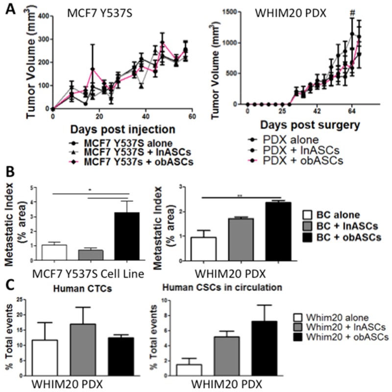
Figure 1.
obASCs promote metastasis but not tumor growth of constitutively active ERα xenograft models—MCF7-Y537S and WHIM20 PDX (A) Tumor volume was tracked over time. (Day of injection = Day 0). There is no change in tumor volume when BC was implanted in the presence of lnASCs or obASCs compared to BC alone except lnASCs compared to control WHIM20 tumor volume at day 60 (# −ln vs. ctrl p < 0.05). Caliper measurements were taken every three to four days until the tumor volume reached 750–1000 mm3. Values reported are the mean (n = 5 mice/group). Data were analyzed using two-way analysis of variance (ANOVA) and a Bonferroni post-test. (B) Area of the lung occupied by metastasis (metastatic index) was evaluated at the endpoint. Groups, where BC was implanted with obASCs, had higher levels of metastasis compared to BC alone or grown with lnASCs. Data were analyzed using one-way ANOVA and Tukey post-test. Bars, ± SEM. * p < 0.05, ** p < 0.01. (C) Circulating tumor cells were analyzed in animals harboring patient-derived xenograft (WHIM20) at endpoint using flow cytometry. There was no change in human (HLA1+) cells across groups; however, analysis of circulating tumor cells enriched for the cancer stem cell marker CD44+CD24− was increased in PDX+obASCs compared to PDX alone. Data were analyzed using one-way ANOVA and Tukey post-test and no significant difference was found. Bars, ± SEM.

