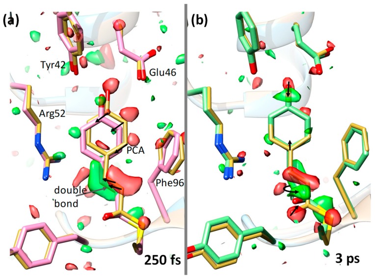
Figure 3.
Ultrafast structural changes in the chromophore pocket of PYP [5] Green represents a positive difference electron density and red a negative difference electron density (on the 3/-3 σ contour level). Yellow structure represents a reference (dark state) structure. The p-coumaric acid (PCA) chromophore as well as some nearby residues are marked. (a) 250 fs after laser excitation (pink structure); the chromophore configuration is still trans. Larger structural changes are denoted by arrows. (b) 3 ps after laser excitation (green structure); the structure is cis. Isomerization occurred about the double bond (curved arrow) at the chromophore tail. Some structural changes are also shown by arrows.

