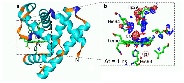
Figure 4.
Time-resolved crystallographic photoflash experiment on the L29W mutant of Mb–CO [66]. (a) Overall structure of MbL29W–CO in the dark. Dashed box: heme pocket. Some important residues are displayed. The heme iron is shown as a yellow sphere. (b) Close-up of the heme pocket 1 ns after an intense optical laser flash to start photodissociation of the CO from the heme. t. The heme and important residues are marked. Red: negative difference electron density; blue: positive difference electron density (−/+ 3 σ contour levels, respectively). Red arrows show structural relaxations at this time delay. In this mutant, the Trp29 transiently occludes the primary docking site of the CO. CO is found at time delays >1 μs on the proximal side of the heme (red-circled p).

