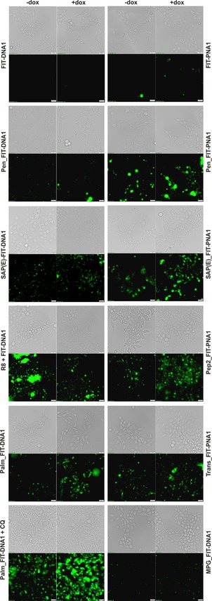
Figure 5.

Fluorescence and bright‐field (gray) microscopy images of living Flp‐In 293 T‐REx cells incubated with DNA (2 μm) or PNA FIT probes (1 μm) for 30 min in PBS at 37 °C and 5 % CO2. Green shows the signal from TO emission with a λ=500/24 nm filter; −dox and +dox: without and with, respectively, the addition of Dox (2 μg mL−1) 1 h before incubation with probes. Scale bar: 20 μm.
