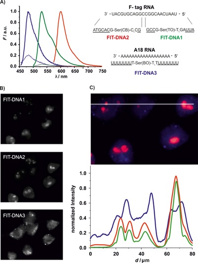
Figure 7.

A) FIT probes for multicolor live‐cell imaging and their emission spectra before (‐ ‐ ‐ ‐) and after (—) addition of target RNA. Conditions: 0.5 μm probe with RNA target (5 equiv) in PBS (100 mm NaCl, 10 mm Na2HPO4, pH 7) at 37 °C. BO: λ ex=440 nm, λ em=485 nm, TO: λ ex=485 nm, λ em=535 nm, QB: λ ex=560 nm, λ em=605 nm, slitex=5 nm, slitem=5 nm. B) Gray‐scale fluorescence microscopy images of Flp‐In 293 T‐REx cells after SLO‐induced delivery of FIT‐DNA1, FIT‐DNA2, and FIT‐DNA3 with induction of F‐tagged mCherry mRNA in separate channels. C) Line scan and normalized intensities for emission from FIT‐DNA1 (green), FIT‐DNA2 (red), and FIT‐DNA3 (blue). Conditions: 2 μg mL−1 Dox; after 1 h incubation with 150 U mL−1 SLO for 10 min in PBS+1 mm MgCl2 and FIT‐DNA1 (2 μm), FIT‐DNA2 (1 μm), and FIT‐DNA3 (4 μm) at 37 °C and 5 % CO2. Filter sets: BO λ ex=438/24 nm, BO λ em=483/32 nm; TO λ ex=500/24 nm, TO λ em=545/40 nm; QB λ ex=575/25, QB λ em=628/40 nm.
