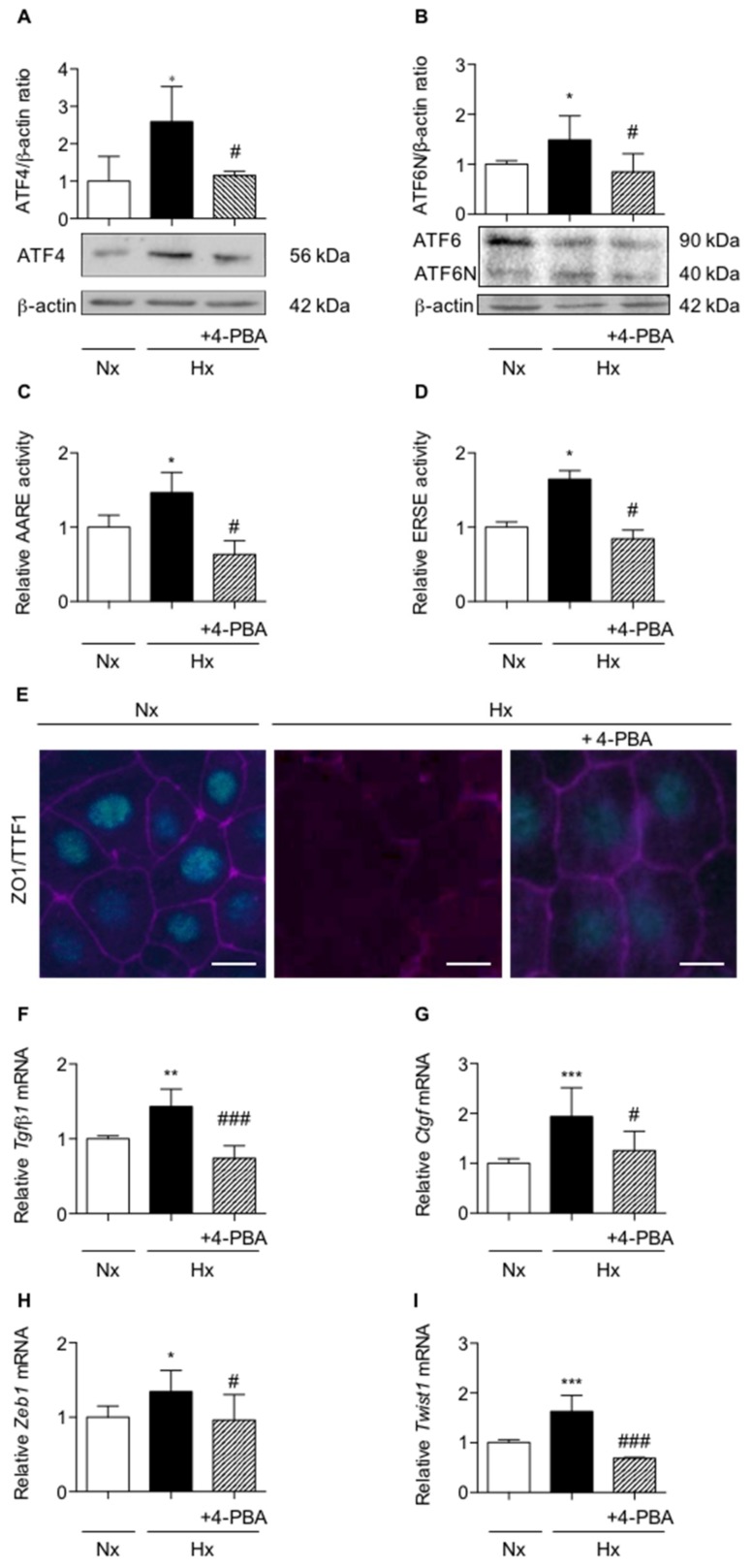
Figure 2.
Inhibition of UPR pathways prevents loss of alveolar epithelial cells phenotype in primary rat alveolar epithelial cells exposed to hypoxia. Isolated primary rat AECs were cultured in normoxia (Nx) (21% O2) or hypoxia (Hx) (1.5% O2) during various periods in the presence or absence of 100 mM 4-phenylbutyrate (4-PBA). (A) Western blot of ATF4 protein and (B) ATF6N/ATF6 ratio were performed primary AECs exposed 6 h to hypoxia. Representative blot of n = 5 experiments is shown. Quantification has been done and the expression levels of ATF4 and ATF6 were reported to the β-actin expression for each condition. (C) Primary rat AECs transfected with plasmid coding for luciferase reporter activity of amino acid response element (AARE: i.e., ATF4-luc) or (D) endoplasmic reticulum stress element (ERSE: i.e., ATF6N/sXBP1-luc), were treated or not with 100 mM 4-PBA and exposed 6 h in hypoxic condition. (C) Luciferase activity corresponding to the transcriptional capacity of ATF4 or (D) ATF6N was measured. (E) ZO-1 (magenta) and TTF1 (cyan) immunostaining were performed on rat AECs cultured on filter and exposed for 6-days to hypoxia in the presence or absence of 100 mM 4-PBA. A representative picture of at least n = 5 independent experiments for each condition has been presented. Scale bar represents 50 µm. (F) mRNA expression levels of Tgf-β1, (G) Ctgf, (H) Zeb1 and (I) Twist1 were quantified by qRT-PCR using 2−∆∆CT method in rat AECs cultured in the presence or absence of 100 mM 4-PBA and exposed 48 h to normoxia or hypoxia. mRNA levels under hypoxic condition were reported to the normoxic condition (n = 5 experiments). Raw data were submitted a Kruskal-Wallis test. *, ** and *** indicate a significant difference as compared with normoxic value with p < 0.05, p < 0.01 and p < 0.001 respectively. # and ### indicate a significant difference as compared with value in untreated hypoxic cells with p < 0.05 and p < 0.001, respectively.

