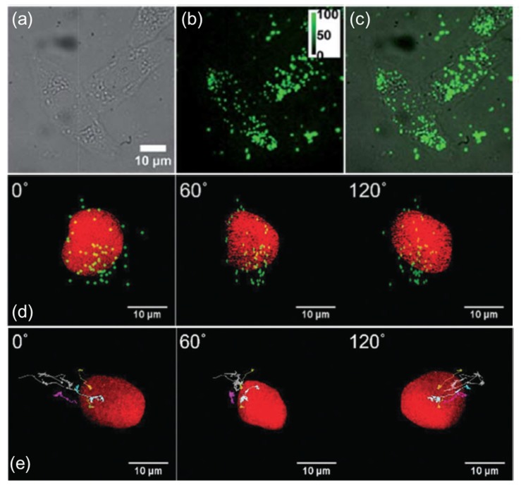
Figure 3.
2D and 3D SPT images. (a–c) The fluorescence microscopy characterization of HeLa cells co-cultured with polymer-modified UCNPs. (a) bright-field, (b) fluorescence and (c) merged microscopy images [90]. (d) The images from various angles (at 0°, 60° and 120°) for UCNP–phospholipid–PEG-TAT. (e) The 3D trajectories from various angles (at 0°, 60° and 120°) for UCNP–phospholipid–PEG-NH3+ [45]. Reproduced from [90] with permission from Royal Society of Chemistry and [45] by permission of the PCCP Owner Societies. Copyright 2018 Royal Society of Chemistry.

