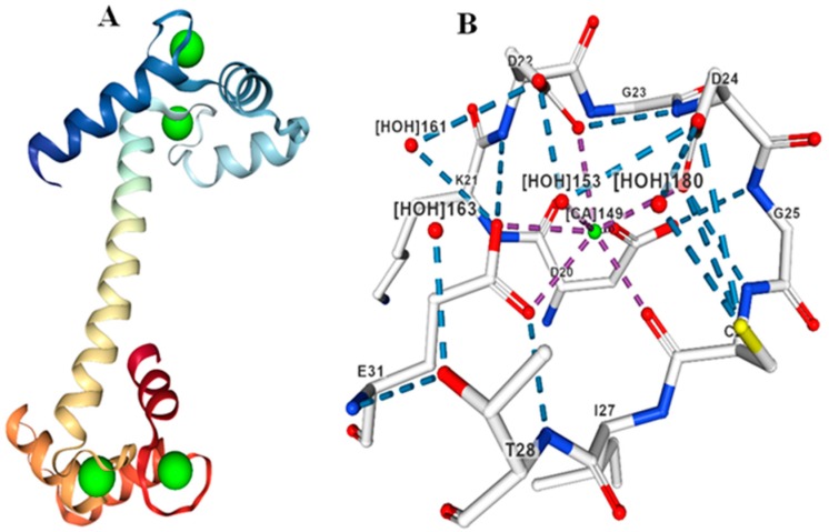
Figure 2.
Molecular structure of CML protein. (A) Three-dimensional structure of CML protein with Ca2+ ligand binding to the EF-hands. (B) D amino acids binding with the Ca2+ ion in the EF-hand of CML protein. Four water molecules (vertices -X) are present adjacent to the Ca2+ ion with hydrogen bonding providing strong structural stability. The molecular structure of CML protein was created according to CML from protein 1UP5 from databank as reported by Rupp et al., (1996) [28].

