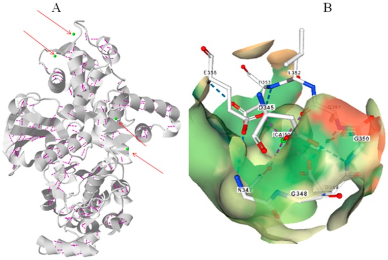
Figure 4.
Molecular structure of CPK protein. (A) Three-dimensional structure of CBL protein with Ca2+ ligands marked in arrow. (B) D-x-D motif binding Ca2+ ion in the EF-hand of CPK protein. No water molecule was found to be associated with the Ca2+ ion in the EF-hand domain of CPK protein. The molecular structure of CPK protein was created according to A. thaliana CPK from protein databank as reported by Chandran et al., (2005) [36].

