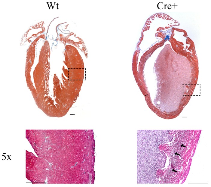
Figure 5.
Histological analysis of 8-month-old Wt and Cre+ hearts. Representative images of 8-month-old wild-type and Cre+ mouse hearts that underwent histological sectioning and staining, imaged under a brightfield microscope. Masson’s trichrome stain was used to visualize the myocardium walls (red) and fibrosis (blue). In addition, 5× magnification using a brightfield microscope was used to identify the presence of fibrosis in Wt or Cre+ hearts, marked with black arrows in fibrotic regions. Scale bar = 500 µm.

