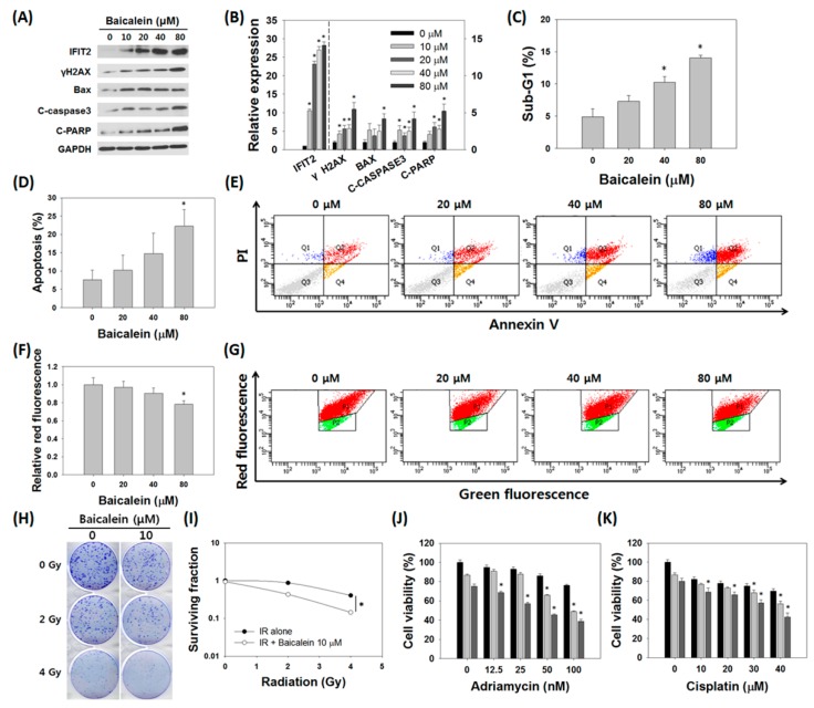
Figure 6.
Baicalein induced apoptosis and sensitized resistant MDA-MB-231/IR cells. (A,B) Expression levels of IFIT2 (left-hand y-axis) and apoptosis marker proteins (right-hand y-axis) after baicalein treatment for 24 h. (C) Cell cycle analysis, (D,E) annexin V/PI staining, and (F,G) JC-1 staining were performed to observe induction of apoptosis by baicalein. (H,I) Baicalein sensitized MDA-MB-231/IR cells to irradiation. (J,K) Baicalein sensitized MDA-MB-231/IR cells to Adriamycin (0, 12.5, 25, 50, and 100 nM) and cisplatin (0, 10, 20, 30, and 40 μM) treatment (▬: Adriamycin or cisplatin alone, ▬: Adriamycin or cisplatin with baicalein 10 μM, ▬: Adriamycin or cisplatin with baicalein 20 μM). Asterisks (*) indicate significant differences at p < 0.05.

