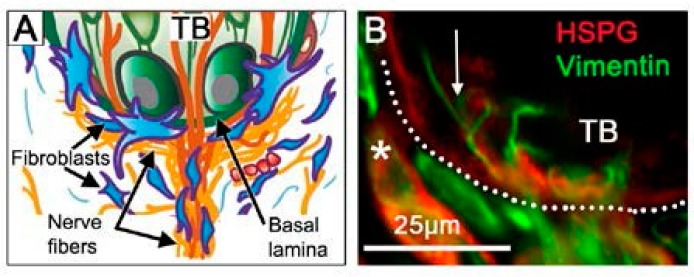
Figure 5.
Basal lamina niche region under the taste bud and at the apex of the fungiform papilla connective tissue core. (A) Diagram for structural interactions among nerve fibers, fibroblasts and lamellipodia, and taste bud (TB) cells, at the basal lamina (Adapted from Figure 4 in Mistretta and Kumari (2017) [13] with permission) (B) Photomicrograph of basal lamina (HSPG immunoreaction, red) under TB cells with lamellipodia extensions (arrow) across the basal lamina from fibroblasts (Vimentin immunoreaction, green). The dotted line demarcates the basal epithelium. The asterisk denotes labelling of blood vessel basal lamina. We propose that Shh is sequestered at the basal lamina in this niche region.

