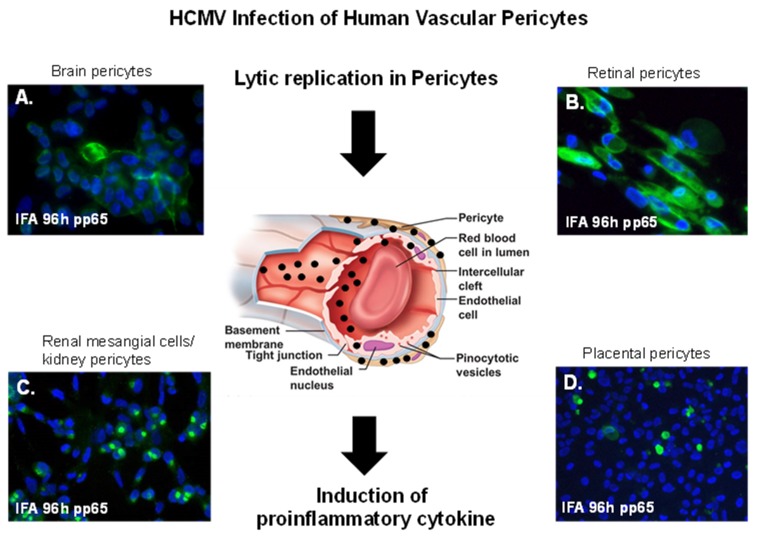
Figure 3.
Vascular pericytes and HCMV infectivity. HCMV lytic infection of human pericytes. Black arrows point to a cross-section of a capillary showing HCMV dissemination in vasculature with virus infection concentrated in pericytes leading to the induction proinflammatory cytokines. (A) Immunofluorescent staining of primary human brain pericytes in with SBCMV 96 h after infection using a mouse monoclonal antibody to the HCMV pp65 tegument protein. (B) Immunostaining of primary human retinal pericytes infected with SBCMV at 96 h stained with the HCMV pp65 antibody. (C) Immunostaining of primary human renal mesangial cells infected with HCMV at 96 h and stained with the HCMV pp65 antibody. (D) Immunostaining of primary human placental pericytes infected with SBCMV at 96 h stained with the HCMV pp65 antibody. All images were taken on a Nikon TE2000S microscope mounted with a CCD camera at ×200 magnification. For fluorescent images, 4′,6-diamidino-2-phenylindole (DAPI) was used to stain the nuclei (blue).

